See where your council stands
See where your council stands
Yolo County bats are no cause for concern.
by Carlos Guerrero [email protected]
May 6, 2020 11:45 am
As the weather is warming, more and more bats will be returning to Yolo County from their winter migration. But is that a cause for concern for the area?
A negative stigma has always followed bats stemming from fictional associations with vampires to nonfictional links to rabies and coronaviruses. A recharged villainization is taking place thanks to the connections between the outbreak of COVID-19 and Chinese Horseshoe bats.
“This villainization of bats is not new, it’s just much worse right now because of the current coronavirus situation,” said Nistara Randhawa, a veterinary epidemiologist, and a postdoctoral scholar at the UC Davis One Health Institute. “The problem comes when we go out and start disturbing spaces that bats live.”

Corky Quirk, a program coordinator for the Yolo Basin Foundation and a volunteer with the wildlife rescue group, Northern California Bats, said that she has been receiving a variety of calls form people that are concerned about whether or not the bats in North America carry the coronavirus.
“I appreciate when people call and ask,” Quirk said. “Sometimes, people are right, and sometimes they are wrong when they jump to their own conclusions, so I’m glad people are looking for more accurate information.”
If a bat is at your home, Quirk suggests gently nudging them at dusk to make them uncomfortable, so they leave, instead of opting to kill or hurt the animal.
According to Quirk, there are around 45 species of bats in the United States, 25 in California, and 17 in northern California.
Around a fifth of all the world’s mammals are bats.
“The DNA of this current virus may be traced to a species of Horseshoe bat in Asia,” Quirk said. “Bats in the United States evolved away from the common ancestor millions and millions of years ago. They are not even closely related. They just happen to be in the same order of animals.”
Woodland buildings are well known for having bats, as are the ones in Davis and West Sacramento. The positives bats provide far outweigh any potential fears over diseases.
“People should really think about what the bat does,” Randhawa said. “The bats are eating pests and insects keeping the insect population down. In Southern California, we have bats that pollinate. They are extremely valuable to the ecosystem.”
Bats may be more likely to get sick from humans than the other way around.
“No, people should not be concerned if they see a bat,” Randhawa said. “The cases are happening because of human to human transmission. The likelihood of humans giving COVID-19 to bats we don’t quite know, but the likelihood of bats giving it to humans is extremely unlikely because these bats have just evolved differently.”
There is such a concern in the other direction that the United States Fish and Wildlife Service has asked scientists to stop bat studies temporarily, for the animal’s safety.
“In California, they are allowing us to take bats into rescue, but they are asking us not to release them once they have healed”, Quirk said. “I’ve had a bat that was trapped in a building in Woodland for a couple of weeks now. Normally I would let her go, but she will stay with me indefinitely until the research is complete as to whether or not humans can make the bats sick.”
As a part of her work with the Yolo Basin Foundation, Quirk is involved in the Bat Talk and Walk event that takes place in June out in the Yolo Bypass Wildlife Area.
Following a 45-minute indoor presentation on bat natural history, a group will carpool out to the Yolo Bypass Wildlife Area to watch the “flyout” of the largest colony of Mexican free-tailed bats in California. The whole experience takes about three hours.
The event looks to be at risk of not happening this year.
First off out of concern for human health, we are waiting to see what the recommendations are for shelter in place,” Quirk said. “We are also discussing how those events could look different if indeed we are or aren’t able to begin to gather again. Whether it’s fewer people or something like that. We’ve looked into having a virtual walk as well.”
According to the Yolo Basin Foundation’s website, the 2020 Bat Talk and Walk session is on hold due to current shelter in place and social distancing recommendations
.”June is pretty far out, but we are trying to figure out alternatives,” Quirk said. “But there is nothing like seeing the animals fly. So I would be sad if we are not able to share that experience. If virtual is the case, then so be it, but we are hopeful that we will be able to share about the bats.”
But if it were to go on as planned, there should be no concern.
Something like the bat walk would be perfectly safe,” Randahawa said. “Your’e not getting close to the bats, and you’re not doing anything that will transfer anything to them.”
by Sandhya Ramesh 24 April 2020
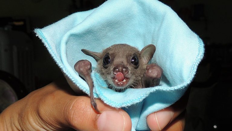
Bengaluru: A group of 64 chiropterologists or bat researchers, scientists, and conservationists from six Asian nations released a statement Friday expressing concern about increased stigmatisation and killing of bats due to “unverified opinions” on these mammals as the source of Covid-19.
The statement was released in response to rising demands by people to kill or remove bats from human neighbourhoods and other natural habitats they occupy.
In the seven-point statement, the chiropterologists have explained how bats haven’t yet been confirmed as the source of the novel coronavirus (the mammals are considered to be the most likely source, but definitive evidence has not been found yet).
They also discuss how bat viruses cannot directly infect humans and there is no evidence that bat faeces can transmit viruses to people.
The statement lists the ecological benefits and vital functions that bats fulfill in terms of pollination and crop protection. It also clarifies that discovery of bat coronaviruses in two species in an Indian Council of Medical Research study poses no health hazard.
The full text of the statement is below:
The world is currently battling a pandemic of unprecedented proportions and bats have been prematurely implicated as the source of COVID-19. Recent social media posts and unverified opinions about bats have led to widespread antipathy and fear in the general public. Incidents of the public requesting for removal of bats, destroying bat roosts, bursting crackers or smoking them out and sealing crevices where bats and their pups roost has increased in the last month both in urban and rural areas in India [1,2]. In this challenging time, we, as people involved in bat conservation at different capacities, would like to clarify that bats do not pose a direct human health hazard. On the contrary, we highlight the role of bats in improving the ecosystem, economy and human health. The following points are listed out in this summary and are elaborated.
1. The exact origin of SARS-CoV-2 or its precursor is not known. It is premature and unfair to blame bats or any other animal for the pandemic.
2. Scientists strongly suggest that it is highly unlikely for SARS-like viruses to jump directly from bats to humans. Also, there is no evidence of humans contracting coronavirus or any such viruses through the excreta of bats .
3. The recent report from the Indian Council of Medical Research (ICMR) on the discovery of bat coronaviruses (BtCoV) in two species of South Asian bats poses no known health hazard. The viruses found in the study are different from SARS-CoV-2 and cannot cause COVID-19.
4. Information on the current, and past zoonotic disease outbreaks suggest that global wildlife trade and/or large-scale industrial livestock farming play an important role in such events. Killing bats and other wild animals, or evicting them from their roosts in retaliation is counterproductive and will not solve any problems.
5. Bats perform vital ecosystem services. They pollinate the flowers of some mangroves, and many other commercially and culturally important plants. Insect-eating bats are voracious eaters of pest insects in rice, corn, cotton and potentially, tea farms.Therefore, bats benefit ecological and human health, and provide intangible economic benefits.
6. The society currently needs more awareness about the bats around them in addition to epidemiological facts for a healthy coexistence. We therefore, request media houses and the press to consider possible negative impacts of their statements on bats and other animals before releasing them.
7. Lastly, we urge the governments of South Asian countries to strengthen the legal framework to protect bats in view of their ecosystem services and their slow breeding capacity.
On the origin and transmission of SARS-CoV-2
The actual origin of SARS-CoV-2 is highly debated among scientists. SARS-CoV-2 appears to be similar to another coronavirus RaTG13 found in a species of bat called the Intermediate Horseshoe Bat (Rhinolophus affinis) [3]. However, a recent study has shown that RaTG13 and SARS-CoV-2 diverged 40-70 years ago from each other (a long timespan for the evolution of viruses) and hence the direct transmission of SARS-CoV-2 or its precursor from bats to humans is improbable [4,5]. Moreover, the surface proteins of all SARS-like viruses found in bats cannot bind efficiently to the corresponding receptors of the human lung epithelium which makes direct transmission even more unlikely [4].
It is also highly improbable that the faeces of bats pose an immediate health risk to humans and, none of the previous zoonotic disease outbreaks, globally, show any evidence that they were caused due to contact with bat faeces [6,7,8]. However, fungal infections (for eg. Histoplasmosis) may arise from unprotected contact with the faeces of any wild animal, so following basic hygiene rules is advised.
Coronaviruses in two species of Indian bats
None of the South Asian bats are proven to be natural reservoirs of SARS-CoV-2. Recently, a study by the Indian Council of Medical Research (ICMR) found bat coronaviruses (BtCoV) in the common Indian Flying Fox (Pteropus medius a.k.a giganteus) and Fulvous Fruit Bat (Rousettus leschenaultii) [9]. However, less than 5% of the screened samples contained this
BtCoV and, as the study mentions, it is very distantly-related to SARS-CoV-2 and hence cannot cause COVID-19.
Relationship between bats and zoonotic disease outbreaks
The real drivers of zoonotic disease outbreaks are predominantly man-made and many animals are carriers of viruses which could potentially spillover to humans. However, in recent times only bats are unfortunately in focus for being reservoirs of viruses. Like any other animal, bats are also reservoirs of many zoonotic viruses. However, being reservoirs does not mean that they spread diseases to humans. In reality, there is very little scientific evidence to prove bats have directly transmitted viruses to humans or caused outbreaks. The only known exception was the Nipah (NiV) outbreak in Bangladesh which was caused through indirect contact between bats and humans. As the cause of the outbreak was identified, it became easy to control and prevent subsequent outbreaks through basic precautionary and mitigation measures [7,10]. Habitat fragmentation, global wildlife trade and wet markets, large-scale industrial farming of wild and domestic animals have synergistic effects in bringing animals in close contact in unsanitary or stressful conditions—ideal conditions for the transmission and evolution of novel zoonotic viruses [11]. Hence, during such outbreaks there is no reason to single out or villainise only bats – whose benefits far outweigh the perceived negativity associated with them. .
Bats are beneficial for humans and the ecosystem
Bats perform vital ecosystem services all over the world. Fruit bats help pollinate globally important cash crops like durian [12] and agave (the plant that produces tequila) [13]. In South Asia, the culturally-significant tree Mahua (Madhuca longifolia) appears to be predominantly bat-pollinated [14]. In Nepal, Chiuri (Diploknema butyracea), a multipurpose tree for the rural populace is pollinated by bats [15]. Small fruit bats also pollinate the flowers of mangroves, which is an extremely productive ecosystem and also a natural barrier to coastal erosion and sea surges, thereby forming our first line of defence to natural disasters. Insectivorous bats, on the other hand, voraciously eat pest insects that cause economic losses in rice plantations [16] and also eat mosquitoes. For instance, bats are estimated to save ~800 million USD for cocoa farmers in Indonesia [17] and ~22 billion dollars (annually) for corn farmers in the United States [18] through pest control. The importance of bats as seed dispersers, pollinators and pest controllers, particularly in the region’s important cash crops (like tea) is beginning to be unravelled. Epidemiologically, the unique immune system of bats could provide clues on handling viruses, therefore they should be seen as a solution to disease outbreaks, rather than the problem.
Concluding remarks
In view of the above points, we firmly believe that the mass hysteria against bats is unfair and uncalled for. The current pandemic is an outcome of the ongoing ecological destruction, increasing intensification of livestock farming and wildlife trade. We urge people not to believe in news from unverified sources and cause harm to bats in retaliation. Likewise, we request the media to not oversimplify scientific evidence, to emphasise the role of humans in disease outbreaks and to highlight the importance of coexistence with bats in urban landscapes. Bats have been living around us for centuries and we have been disease free wherever bats have been left to their business. Oversimplified or unverified information from the press not only creates unnecessary fear among the public but also pushes decades of conservation efforts backwards which is far more destructive for the ecosystem.. In India, only two species (out of 128) are protected by law, while many other species are more endangered or lack scientific information [19]. In Nepal, too, all species are unprotected, including two species from the National Red List [20] and the same is true for Pakistan. We urge the governments of these countries to reconsider and reinforce the laws governing bat conservation.
Signatories
*Email IDs are provided for those who have agreed to be contacted by the media
1. Rohit Chakravarty, Leibniz Institute for Zoo and Wildlife Research, Berlin, Germany <[email protected]>
2. Baheerathan Murugavel, Indian Institute of Science Education and Research Thiruvananthapuram (IISER TVM), India <[email protected]>
3. Dr. Seshadri K S, Indian Institute of Science (IISc) and The Madras Crocodile Bank Trust (MCBT), India <[email protected]>
4. Vidisha Kulkarni, Jain University and GubbiLabs, Bangalore, India
5. Rajesh Puttaswamaiah, Citizen Scientist & Trustee, Bat Conservation India Trust, Bangalore, India <[email protected]>
6. Dr. Vadamalai Elangovan, Babasaheb Bhimrao Ambedkar University, Lucknow, Uttar Pradesh, India <[email protected]>
7. Shriranjani Iyer, Sálim Ali Centre for Ornithology and Natural History (SACON), Anaikatty, Coimbatore, India <[email protected]>
8. Aditya Srinivasulu, Biodiversity Research and Conservation Society, Hyderabad, India. <[email protected]>
9. Dr. Pushpa Raj Acharya, Central Campus of Science and Technology, Mid-Western University, Birendranagar, Surkhet, Nepal <[email protected]>
10. Basanta Sharma, Nepal Bats Research and Conservation Union (NeBRCU), Pokhara, Nepal. <[email protected]>
11. Dr. T.Ganesh, Senior Fellow, Ashoka Trust for research in Ecology and the Environment(ATREE), Bangalore, India
12. Sanjeev Baniya, Nepal Bat Research and Conservation Union (NeBRCU), Pokhara, Nepal. <[email protected]>
13. Dr. A. Rathinakumar, Madurai Kamaraj University, Madurai, India. <[email protected]>
14. Dr. Chetan HC, Assistant Professor, The University of Transdisciplinary Health Sciences and Technology, Centre for Conservation of Natural resources, Bengaluru, India <[email protected]>
15. Dr. R. Ganesan, Ashoka Trust for Research in Ecology and the Environment (ATREE), Bangalore, India
16. Rohit Chouhan, Wildlife Research Fellow, Department of Wildlife Science, University of Kota, Kota, Rajasthan, India. <[email protected]>
17. Kaushik N, Madurai Kamaraj University, Madurai, India <[email protected]>
18. Dr. Sumit Dookia, Assistant Professor, University School of Environment Management, Guru Gobind Singh Indraprastha University, New Delhi, India <[email protected]>
19. Aishanya Sarma, Conservation Initiatives, Assam, India <[email protected]>
20. Kasturi Saha, Indian Institute of Science (IISc), Bangalore, India <[email protected]>
21. Ram Mohan, Indian Institute of Science Education and Research Pune (IISER Pune), India <[email protected]>
22. Tariq Ahmed Shah, Wildlife Biology & Taxonomy Lab, Osmania University, Hyderabad, India. <[email protected]>
23. Devna Arora, Rehabber’s Den, Pune, India <[email protected]>
24. Harish Prakash, Indian Institute of Science (IISc), Bangalore, India <[email protected]>
25. Dr. Parvathy Venugopal, School of Biological Sciences, University of Bristol, United Kingdom. <[email protected]>
26. Rajlakshmi Mishra, University School of Environmental Management, Guru Gobind Singh Indraprastha University, New Delhi, India <[email protected]>
27. Rahul Prabhukhanolkar, Mhadei Research Center, Belagavi and Indian Bat Conservation Research Unit, India <[email protected]>
28. A. Karthikeyan, Madurai Kamaraj University, Madurai, India. <[email protected]>
29. Prof. Hema Somanathan, Indian Institute of Science Education and Research, Thiruvananthapuram (IISER-TVM), India <[email protected]>
30. Prof. G. Marimuthu, Madurai Kamaraj University, Madurai, India
31. Dr. Utttam Saikia, Zoological Survey of India, Shillong, India <[email protected]>
32. Dr. H. Raghuram, PG and Research Department of Zoology, The American College, Madurai 625 002, Tamil Nadu, India <[email protected]>
33. Tharaka Kusuminda, Department of Agricultural Biology, Faculty of Agriculture, University of Ruhuna, Kamburupitiya, Sri Lanka. <[email protected]>
34. Jayanthi Kallam, Founder and Executive Director, Avian and Reptile Rehabilitation Trust, Bangalore, India <[email protected]>
35. Prof. Sripathi Kandula, Madurai Kamaraj University / Chettinad Academy of Research and Education, Chennai, India <[email protected]>
36. Dr. Amani Mannakkara, Department of Agricultural Biology, Faculty of Agriculture, University of Ruhuna, Kamburupitiya, Sri Lanka. <[email protected]>
37. Ravi Umadi, Department of Biologie II, Biocenter, Ludwig-Maximillians Universitaet, Munich, Germany <[email protected]>
38. Sargam Singh Rasaily IFS, APCCF/Member Secretary, Uttarakhand Biodiversity Board, Dehradun, Uttarakhand, India <[email protected]>
39. R.M.T. Priyanwada Rathnayake, Master of Environment Management , Faculty of Graduate Studies , University of Colombo, Sri Lanka <[email protected]>
40. Sangay Tshering, College of Natural Resources, Royal University of Bhutan, Punakha, Bhutan. <[email protected]>
41. Dr. D. Paramanantha Swami Doss, Assistant Professor, St. John’s College, Palayamkottai, Tamil Nadu, India <[email protected]>
42. Dr. Venkatesh Nagarajan Radha, Postdoc Associate, University of Sydney, Australia. <[email protected]>
43. Dr. Chelmala Srinivasulu, Department of Zoology, Osmania University, Hyderabad, Telangana State, India <[email protected]>
44. Dr. Bhargavi Srinivasulu, Department of Zoology, Osmania University, Hyderabad, Telangana State, India
45. Dr. S. Baskaran, Assistant Professor, Department of Biotechnology, The Madura College, Madurai, Tamil Nadu, India <[email protected]>
46. Chamara Amarasinghe, Faculty of Graduate Studies, University of Colombo, Sri Lanka. <[email protected]>
47. Pratik Das, XVI M.Sc., Wildlife Institute of India, Dehradun, India.
48. Dr. Touseef Ahmed, Department of Biological Sciences, Texas Tech University, Texas, USA. <[email protected]>
49. Dr. T. Karuppudurai, Madurai Kamaraj University, Madurai, India. <[email protected]>
50. Steffi Christiane R, Department of Animal Behaviour & Physiology, School of Biological Sciences, Madurai Kamaraj University, Madurai, India. <[email protected]>
51. Dr. Manjari Jain, Indian Institute of Science Education and Research, Mohali (IISER-M), India <[email protected]>
52. Suranjan Karunarathna, Nature Exploration and Education Team, Colombo, Sri Lanka <[email protected]>
53. Dr. Sanjay Molur, Co-chair, Chiroptera Conservation & Information Network of South Asia (CCINSA), Zoo Outreach Organization, Coimbatore, India. <[email protected]>
54. Dr. Kranti Yardi, Professor, Bharati Vidyapeeth Institute of Environment Education and Research, Bharati Vidyapeeth Deemed to be University, Pune <[email protected]>
55. Aita Hang Subba, Guest faculty, Department of Zoology, Sikkim University, Gangtok, India <[email protected]>
56. M. Mathivanan, Senior Research Associate, Ashoka Trust for Research in Ecology and the Environment (ATREE), Agasthyamalai Community Conservation Centre (ACCC), Manimutharu, Tirunelveli, Tamil Nadu, India <[email protected]>
57. Dr. Adora Thabah, Freelance researcher, Shillong, Meghalaya. <[email protected]>
58. Tijo K Joy, UNDP Cluster Coordinator- Conservation & Ecology HTML Project, Munnar, Kerala, India.
59. Dr. K. Emmanuvel Rajan, Department of Animal Science, School of Life Sciences, Bharathidasan University, Tiruchirappalli-620024, Tamil Nadu, India <[email protected]>
60. Shasank Ongole, National Centre for Biological Sciences, Bengaluru, Karnataka
61. Soham Mukherjee, Herpetologist & Wildlife Biologist, NAJA India, Ahmedabad (Gujarat), India <[email protected]>
62. Kadambari Deshpande, Ashoka Trust for Research in Ecology and the Environment (ATREE), Bangalore, India
63. Thejasvi Beleyur, Max Planck Institute for Ornithology, Seewiesen, Germany <[email protected]>
64. Dr Md Nurul Islam, FETPV Technical Officer, Global Health Development (GHD), Bangladesh <[email protected]>
Note: The views of the signatories are personal and may not reflect those of their institutions.
References
1. “Bats are Bengaluru’s enemy no 1 now”, news article in Bangalore Mirror on 20th
April 2020. https://bangaloremirror.indiatimes.com/bangalore/cover-story/bats-are-
bengalurus-enemy-no-1-now/articleshow/75240633.cms
2. “Myths of bats spreadinng Coronavirus: Two trees chopped off in city”, news report
in Star of Mysore on 3rd April 2020. https://starofmysore.com/myth-of-bats-
spreading-coronavirus-two-trees-chopped-off-in-city/
3. Zhou, P., Yang, X., Wang, X. et al. (2020). A pneumonia outbreak associated with a
new coronavirus of probable bat origin. Nature 579, 270–273 (2020).
https://doi.org/10.1038/s41586-020-2012-7
4. Andersen, K.G., Rambaut, A., Lipkin, W.I. et al. (2020) The proximal origin of
SARS-CoV-2. Nat Med 26, 450–452. https://doi.org/10.1038/s41591-020-0820-9
5. Boni, M.F., Lemey, P., Jiang, X. et al. (2020). Evolutionary origins of the SARS-
CoV-2 sarbecovirus lineage responsible for the COVID-19 pandemic. Preprint on
www.biorxiv.org. https://doi.org/10.1101/2020.03.30.015008
6. World Health Organization Fact Sheet on Ebola: https://www.who.int/news-
room/fact-sheets/detail/ebola-virus-disease
7. World Health Organization Fact Sheet on Nipah virus: https://www.who.int/news-
room/fact-sheets/detail/nipah-virus
8. World Health Organization Fact Sheet on Middle Eastern Respiratory Syndrome:
https://www.who.int/news-room/fact-sheets/detail/middle-east-respiratory-syndrome-
coronavirus-(mers-cov)
9. Yadav, P.D, Shete-Aich, A., Nyayanit, D.A., et al. (2020). Detection of coronaviruses
in Pteropus and Rousettus species of bats from different states of India. Indian
Journal of Medical Research
10. Dhillon, J., Banerjee, A. (2015) Controlling Nipah virus encephalitis in Bangladesh:
Policy options. J Public Health Pol 36, 270–282.
https://doi.org/10.1057/jphp.2015.13
11. Jones, B.A, Grace, D., Kock, R. et al. (2013). Zoonosis emergence linked to
agricultural intensification and environmental change. PNAS 110 (21), 8399-8404.
https://doi.org/10.1073/pnas.1208059110
12. Aziz, SA, Clements, GR, McConkey, KR, et al. (2017) Pollination by the locally
endangered island flying fox (Pteropus hypomelanus) enhances fruit production of the
economically important durian (Durio zibethinus). Ecol Evol.; 7: 8670– 8684.
https://doi.org/10.1002/ece3.3213
13. Trejo-Salazar, R.E, Eguiarte, L.E, Suro-Piñera, D. and Medellin, R.A. (2016) Save
Our Bats, Save Our Tequila: Industry and Science Join Forces to Help Bats and
Agaves Natural Areas Journal 36(4), 523-530. https://doi.org/10.3375/043.036.0417
14. Nathan, P.T., Karupuddurai, T., Raghuram, H. and Marimuthu, G. (2009). Bat
foraging strategies and pollination of Madhuca longifolia (Sapotaceae) in southern
India. Acta Chiropterologica, 11(2): 435-441.
15. Acharya, P.R. (2015). Chepang Chiuri and Chamera. Friends of Nature, Kathmandu.
16. Wanger, T.C., Darras, K., Bumrungsri, S. et al. (2014). Bat pest control contributes to food security in Thailand. Biological Conservation, 171: 220-223.
17. Maas, B., Clough, Y. and Tscharntke, T. (2013). Bats and birds increase crop yield in
tropical agroforestry landscapes. Ecology Letters, 16: 1480-1487.
18. Maine, J. and Boyles, J.G. (2015). Bats initiate vital agroecological interactions in
corn. PNAS, 112(4): 12438-12443.
19. Srinivasulu, C., Srinivasulu, A. and Srinivasulu. B. (2020). Checklist of the bats of
South Asia (v1.1). https://threatenedtaxa.org/index.php/JoTT/checklists/bats/southasia
[Date of publication: 13 April 2020].
20. Jnawali, S.R., Baral, H.S., Acharya, K.P., Upadhyay, G.P. et al. (2011). The Status of
Nepal Mammals: The National Red List Series, Department of National Parks and
Wildlife Conservation, Kathmandu, Nepal.
Also read: Bat coronavirus found in two Indian species of bats for the first time: ICMR study
AUSTIN BATS and COVID-19:
• Bats in Austin do not have or spread SARS-CoV-2 (the virus that causes COVID-19 in humans). SARSCoV-2 is not found in North American bat species at present.
• Transmission of COVID-19 is from humans to other humans.
• There are theoretical concerns about the possibility for transmission of SARS-CoV-2 from humans to bats. Research is underway; until we know more, precautions to minimize the chance of North American bats of being exposed to SARS-CoV-2 are being taken. This is not unique to bats – there are also concerns of possible transmission from humans to other wildlife species, particularly mustelids, felids and canids.
• Avoid handling live bats and any wildlife. If handling live bats cannot be avoided, follow recommendations about PPE to prevent spread of respiratory droplets – a face mask, thick gloves, etc.
Bat conservation and COVID-19
• Many bat species have adapted to urban and rural environments, where they coexist safely with people. Ask us about living with bats safely.
• It is important that bats and bat habitat not be destroyed because of unfounded fears over coronavirus transmission.
• Bats in North America are in trouble from habitat loss and white-nose syndrome – at least two species in Texas have experienced sharp decline just this spring.
• Bats are essential to our Texas ecosystems and economy. Nationwide, bats are estimated to provide $23 billion of natural pest control each year.
• Killing bats would not have any effect on the spread of COVID-19, but would negatively affect bat populations, conservation efforts, and our economy.
Austin Bat Refuge would like to remind people we are here to answer your questions and collect reports on bat sightings, bat roosts, and dead bats. Contact us at [email protected] and 512-695-4116 or 512-799-8847.
(This post borrowed and modified from BC Community Bat Program)
Ever wonder about bat’s pest control services? We see evidence every morning that bats are at work for us, all night long.
We grow crops to attract moths; the females lay their eggs on our brassicas such as collard greens, curly kale, swiss chard, and cabbage. We allow the larvae to eat all they want of the nutritious greens and they soon become moths. Without bats to balance the numbers of crop pests, our food crops would take a beating.
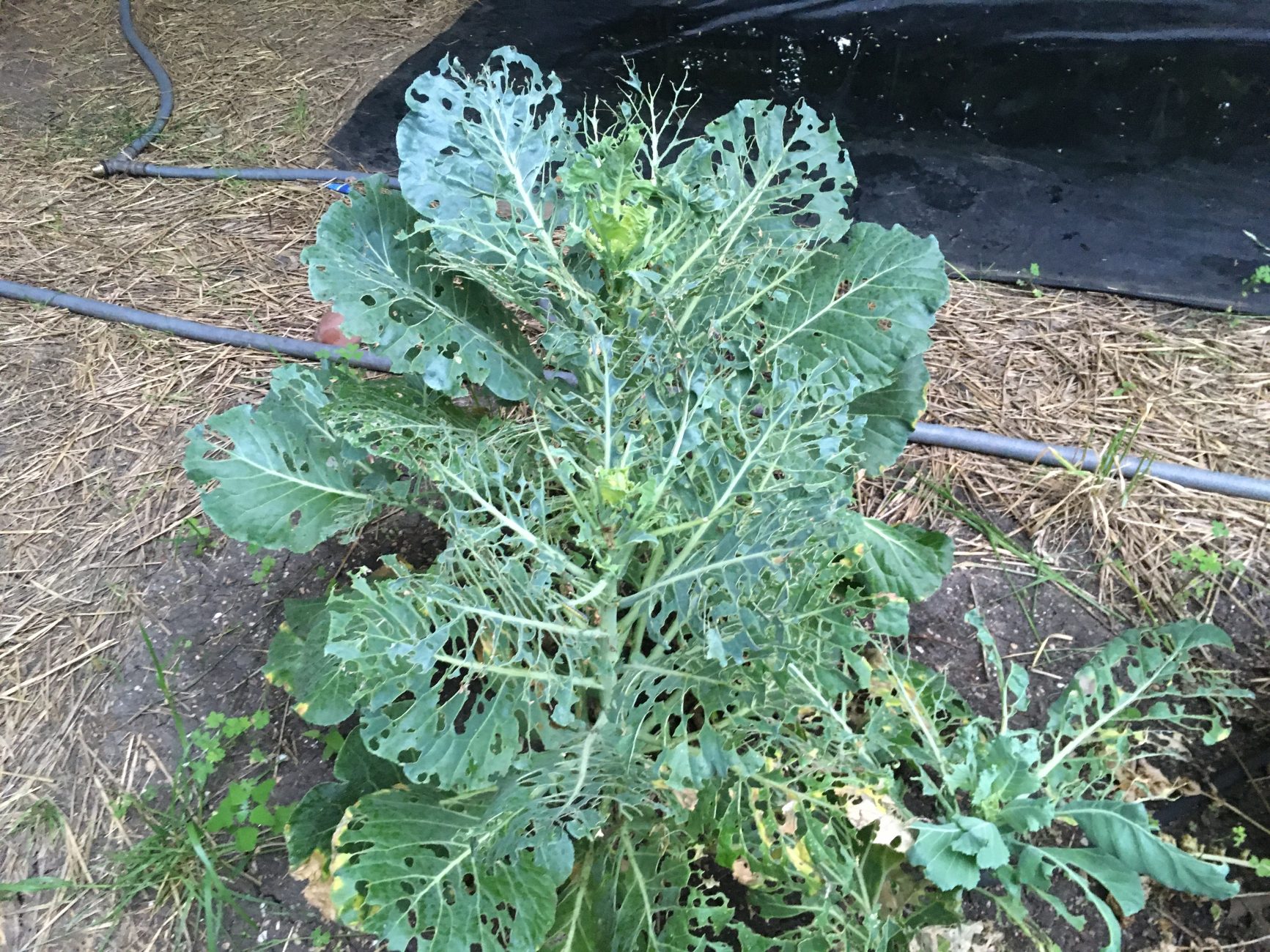
The flight cage is full of moths nowadays and the rehabilitating bats are loving their hunting practice.
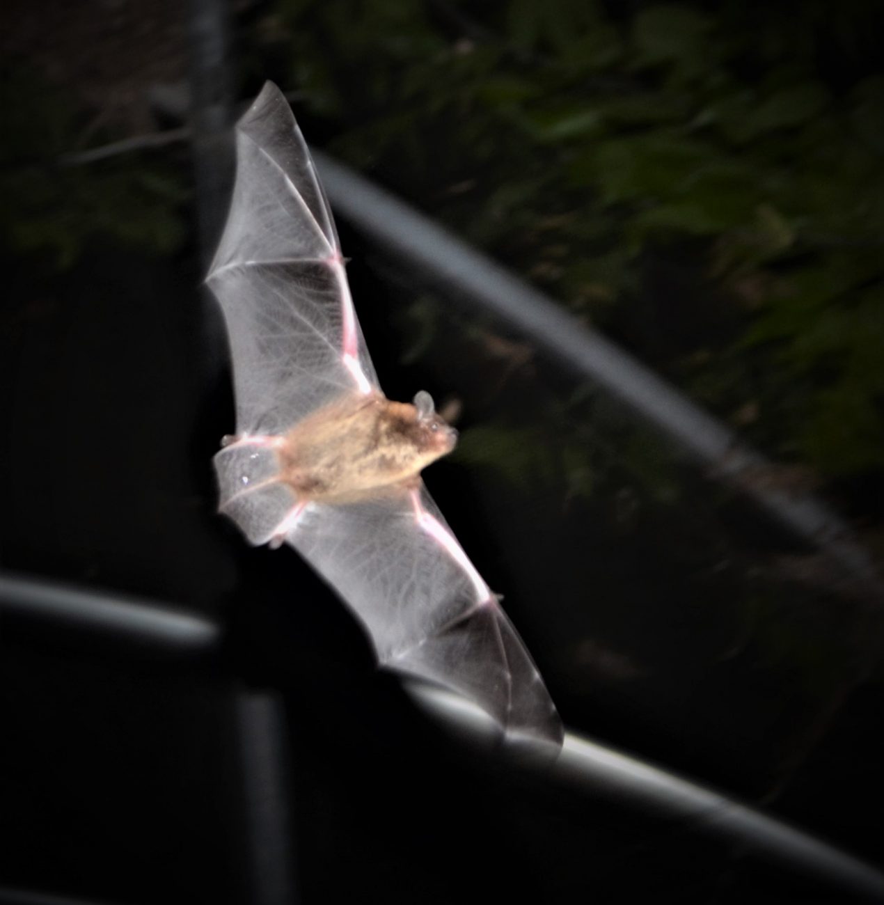

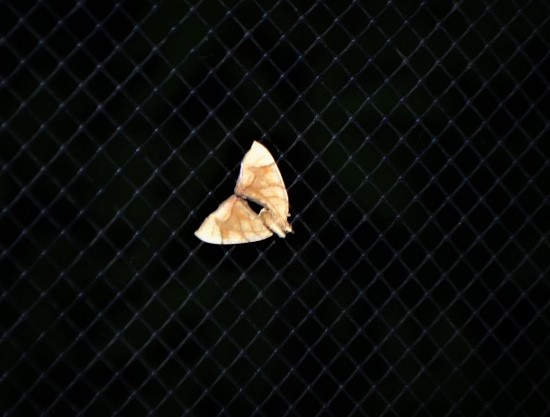
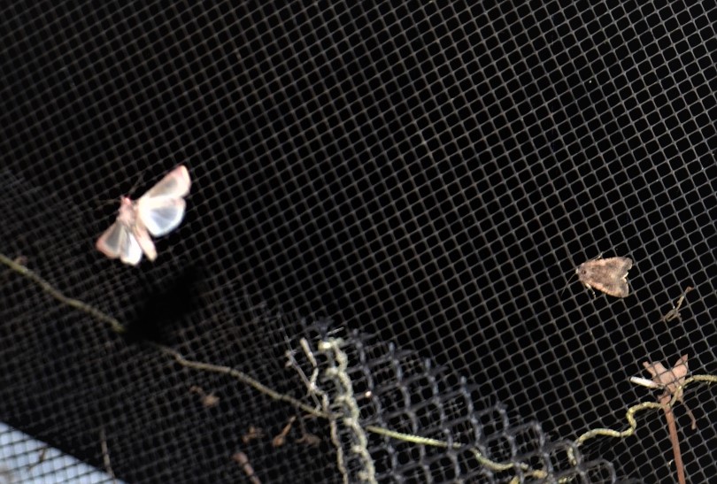
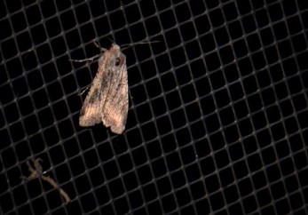

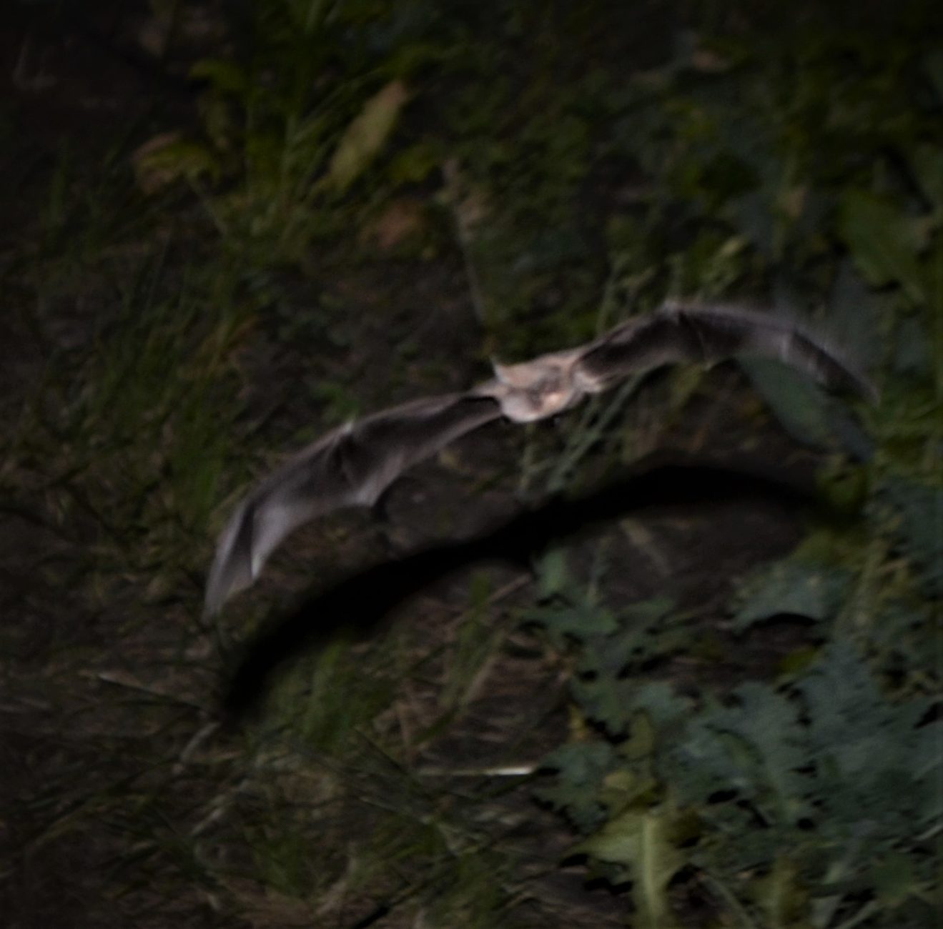
When moths hear echolocation of an approaching bat, millions of years of evolution kick in and they employ various defense mechanisms. In our aviary some fly erratically while others drop to the ground to evade the bats. Normally dropping works well as they hide in the grass, but in our fight cage they often drop into the bats’ drinking troughs. Each morning we go out to rescue the moths trapped by the surface tension of the water.
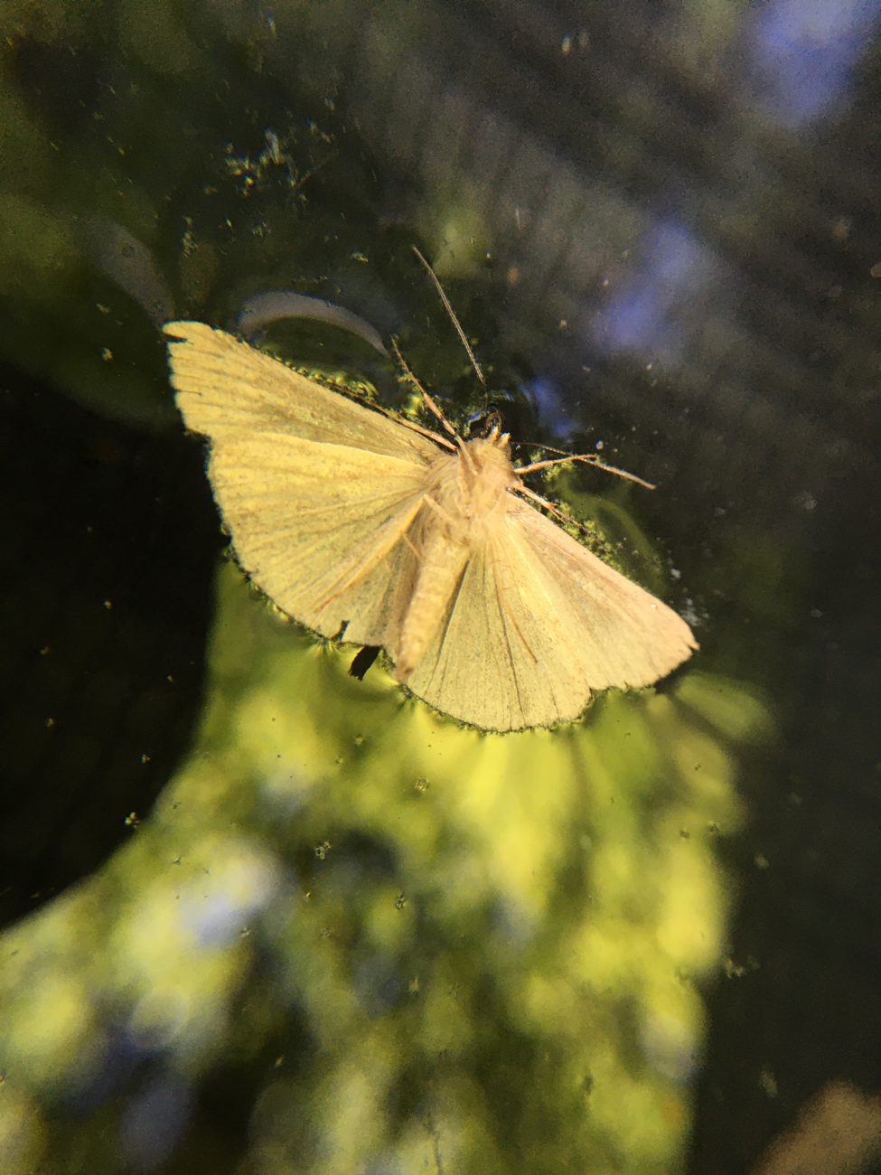
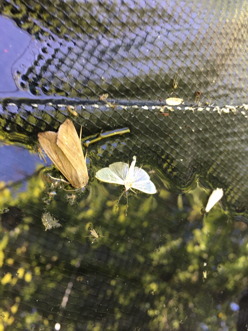
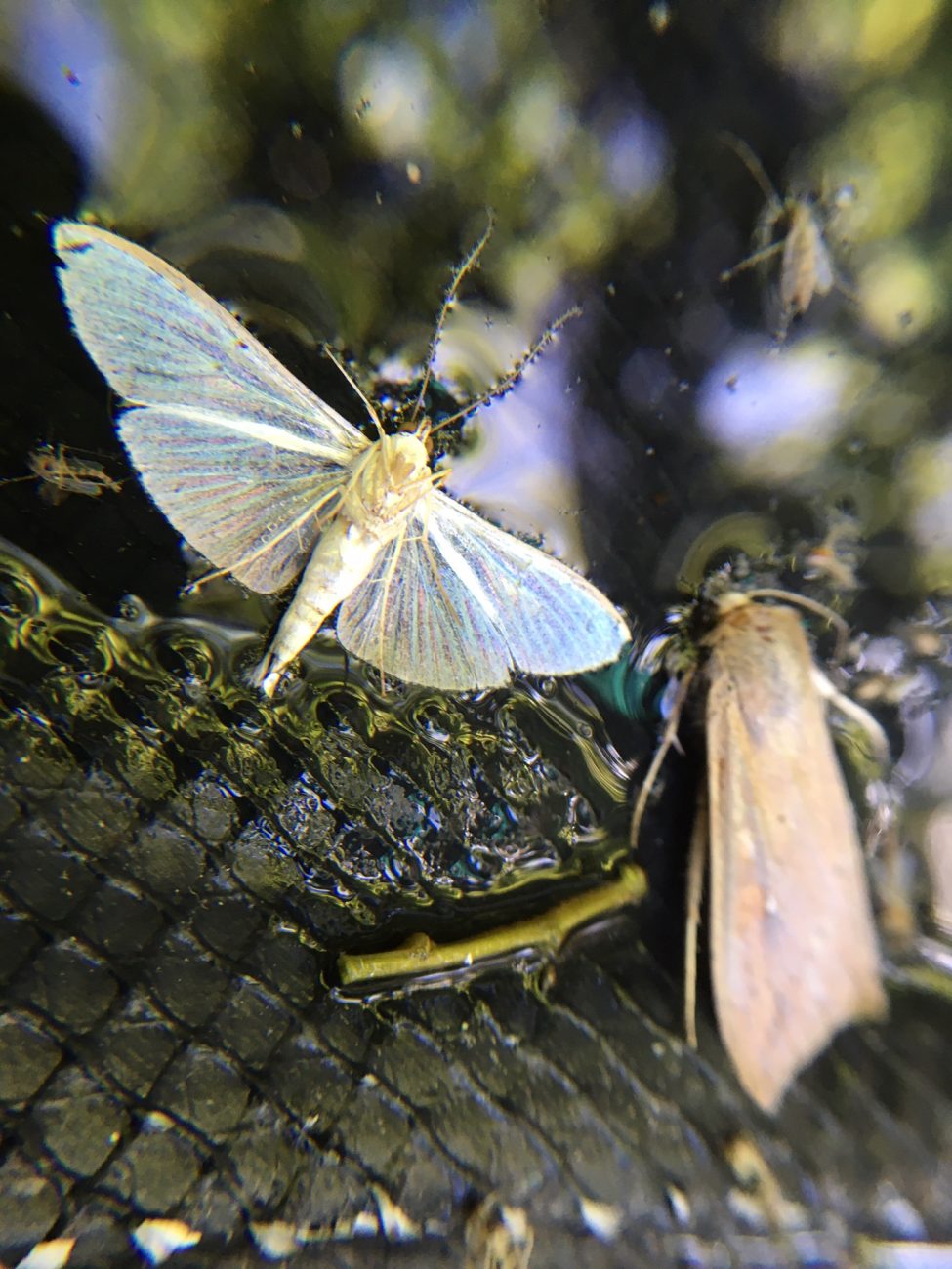


Soon the brassicas will have served their purpose. We’ve eaten lots this winter, but as the insects begin to ravage them in the spring, they form a bitter latex in their leaves that makes them unpalatable to the larvae, but also to us! Soon we will replace them with a cover crop, to fix nitrogen in the soil and protect the soil from the hot summer sun.
And we’ll plant the winter garden again in the fall.
Cycle of life.
By Dr. Tigga Kingston | Originally published April 17, 2020
Bats have earned an unwarranted reputation as disease spreaders since the Covid-19 outbreak. With April 17 marking World Bat Appreciation Day, Dr. Tigga Kingston sets out to provide the full picture on these misunderstood mammals.
It’s 7:20pm at the edge of the rainforest of Krau Wildlife Reserve in Peninsular Malaysia. Plaintive calls of nightjars herald the transition from day to night as silhouettes of bats flitter in the twilight.
Bats do not spread Covid-19 — you can only catch it from other people. SARS-CoV-2 is the virus that causes Covid-19. A related virus, RatG2013, was isolated from an intermediate horseshoe bat from China in 2013, leading to suggestions the bat virus jumped to people. Research shows this is extremely unlikely. The key part of the virus that enables it to infect people is not from bats — the closest match identified is from a pangolin.
What is becoming clear is that human activities are making viral spillover events more likely. Habitat disturbance stresses animals, making them more susceptible to viruses and more likely to produce high viral loads. Humans are encroaching on wildlife habitat and trading or consuming wildlife, increasing exposure opportunities.
Bats provide enormous ecological and economic value worldwide. They are important pollinators and seed dispersers for hundreds of plants. Most famously, they are the key pollinator of durian — an 18 billion dollar industry. No bats means no durian.
The wrinkle-lipped free-tailed bat roosts in caves in the thousands and millions. One colony in Thailand of 2.6 million bats eats about four tonnes of plant-hoppers — a major rice pest — in a single night. Across Thailand this one species saves about 2,892 tonnes of rice annually.
Loss of habitat is a major challenge facing bats globally. About half of Southeast Asia’s bats roost in caves that are being lost to tourism disturbance, guano harvesting, and limestone quarrying for the cement industry. Intense hunting of large fruit bats is pushing many species toward extinction.
The solutions are familiar: habitats must be protected and restored, wildlife exploitation must stop, and the world’s ecosystems must be kept healthy. Creating buffers between human settlements and wildlife is essential for protecting both public and environmental health.
Bats are long-lived mammals — some species live more than 30 years — that typically give birth to only one pup per year. Their populations cannot withstand sustained disturbance or intense hunting. Every bat that survives is important.
By Melissa Ingala & Ariadna Morales | Article published in Global Bat Net, April 20, 2020
Many people assume all bats carry diseases threatening humans. This concern intensified after a virus from one bat species appeared related to SARS-CoV-2. Research indicates bats serve as special reservoirs for zoonotic pathogens due to their flight ability, immune systems, and colonial behavior.
However, recent studies demonstrate that bats are no more likely to transmit zoonotic viruses than other groups of mammals when accounting for species diversity. The abundance of viruses found in bats likely reflects the sheer number of bat species — over 1,400 — rather than any inherent danger.
Culling bat populations does not prevent disease transmission and may actually backfire. Vampire bat removal in Peru increased rabies transmission among remaining populations, which skewed toward juveniles with less immunity. Endangered bats show higher viral loads than stable populations, suggesting that extinction threats can worsen viral hosting capacity.
Bats provide enormous ecological and economic value. They deliver millions of dollars in pest control services globally and pollinate crops including tequila agaves, mangos, and durian. They also combat deforestation and climate change through seed dispersal and rainforest regeneration.
Preventing future spillovers requires behavioral change and habitat protection. Education about avoiding contact with disease reservoirs is effective. Deforestation and human encroachment in Africa correlate with Ebola-positive fruit bats. Creating buffers between human settlements and wildlife is essential for protecting both public and environmental health.
The answer is not to harm bats — it is to protect them and the habitats that keep our ecosystems, and ourselves, healthy.
View ORCID ProfileNardus Mollentze and View ORCID ProfileDaniel G. Streicker PNAS first published April 13, 2020 https://doi.org/10.1073/pnas.1919176117
Identifying whether novel human viruses disproportionately originate from certain animal groups could inform risk-based allocations of research and surveillance effort. Whether such “special reservoirs” exist remains controversial. We show that the proportion of viruses that infect humans varies minimally across reservoir taxonomic orders. Instead, the number of human-infecting viruses increases proportionately to the total number of viruses maintained by each reservoir group, which is in turn explained by the number of animal species within each group. This supports a host-neutral explanation for observed variation in the number of zoonoses among animal groups, such that traits of animal orders are unlikely to produce viruses that disproportionately threaten humans. These findings refine strategies to identify high-risk viruses prior to their emergence.
The notion that certain animal groups disproportionately maintain and transmit viruses to humans due to broad-scale differences in ecology, life history, and physiology currently influences global health surveillance and research in disease ecology, virology, and immunology. To directly test whether such “special reservoirs” of zoonoses exist, we used literature searches to construct the largest existing dataset of virus–reservoir relationships, consisting of the avian and mammalian reservoir hosts of 415 RNA and DNA viruses along with their histories of human infection. Reservoir host effects on the propensity of viruses to have been reported as infecting humans were rare and when present were restricted to one or two viral families. The data instead support a largely host-neutral explanation for the distribution of human-infecting viruses across the animal orders studied. After controlling for higher baseline viral richness in mammals versus birds, the observed number of zoonoses per animal order increased as a function of their species richness. Animal orders of established importance as zoonotic reservoirs including bats and rodents were unexceptional, maintaining numbers of zoonoses that closely matched expectations for mammalian groups of their size. Our findings show that variation in the frequency of zoonoses among animal orders can be explained without invoking special ecological or immunological relationships between hosts and viruses, pointing to a need to reconsider current approaches aimed at finding and predicting novel zoonoses.
Most emerging infectious diseases of humans are viruses that originate from nonhuman animals via “zoonotic” transmission (1⇓–3). Given the diversity of animals and viruses in nature, targeting virus discovery, surveillance, and research toward the taxonomic groups with the highest propensity to infect humans would benefit attempts to mitigate future disease outbreaks (4⇓⇓–7). Identifying these groups remains a major challenge. Viruses with RNA genomes are overrepresented as zoonoses and certain families including Filoviridae, Orthomyxoviridae, and Togaviridae contain a large proportion of zoonotic species (8, 9). Among hosts, however, patterns are less clear. Large-scale comparative studies have suggested that barriers to cross-species transmission increase with evolutionary divergence from humans, implying heightened zoonotic risk from closely related nonhuman primates (9, 10). Yet, other animal groups including bats, rodents, and ungulates are also frequently associated with zoonoses despite their evolutionary distance from humans (9, 11⇓–13). A popular explanation (here, the “special reservoir hypothesis”) is that physiological or ecological traits of these taxa make them more likely to maintain zoonotic viruses or transmit them to humans. For example, ungulates and rodents include domesticated or anthropophilic species whose high ecological overlap with humans could facilitate pathogen exchange (13, 14). In contrast, the unique life history of bats has been hypothesized to create an evolutionarily distinct immunological environment that selects for viral traits that favor human infection (15⇓⇓⇓⇓–20). An alternative to the special reservoir hypothesis is that host species maintain a similar number of viruses with a similar per-virus risk of zoonotic transmission. Variation in the number of zoonoses among animal groups therefore arises as a consequence of their species richness (here, the “reservoir richness hypothesis”). For example, the preponderance of rodent- or bat-associated zoonoses could reflect the large number of rodent and bat species relative to other mammalian groups (21). These hypotheses imply different management strategies. The special reservoir hypothesis would advocate fundamental research to define the ecological or physiological trait profiles that explain the propensity of certain hosts to harbor zoonoses, followed by targeted surveillance or virus discovery in hosts with high-risk trait profiles. In contrast, the reservoir richness hypothesis would advocate broader surveillance and virus discovery, possibly proportionate to the local species richness of different animal groups, and would imply the need for deeper understanding of which features of virus biology enhance zoonotic transmission.
Identifying patterns in the animal origins of zoonoses has been frustrated by disconnects between the key outcome of interest, the likelihood of zoonotic transmission, and currently available data which document viral infection in human and nonhuman hosts but provide limited information on the origins of these infections. For example, humans may infect nonhuman hosts, rather than the other way around, but only the latter direction of transmission is pertinent to zoonotic origins. A greater challenge is that the reservoir host species that maintain and transmit viruses to humans can rarely be distinguished from species that form a larger community of “dead-end” hosts that are inconsequential to transmission (22, 23). Rather than reflecting genuine imbalances in the animal origins of human infections, associations between certain animal groups and zoonotic viruses based on shared detections might instead emerge from heightened surveillance for or susceptibility to zoonotic viruses in these groups even if they are not key components of natural transmission cycles. As the known diversity of viruses expands, the increasing difficulty of differentiating related viruses in different host species from the same virus transmitted between these host species would exacerbate this effect by overestimating viral sharing, particularly if relying on serological or PCR data alone (24).
Given this uncertainty in the reservoir hosts of most viruses at the species level, an alternative approach to resolving whether animal host associations influence zoonotic risk would be to assess reservoir associations at lower taxonomic resolutions. Such analyses would take advantage of the tendency for viruses to be maintained by host species within the same taxonomic order, a phenomenon which arises from more frequent host shifting among closely related species and the cospeciation of hosts and viruses (25⇓–27). At this coarser taxonomic level, transmission cycles may be accurately defined for hundreds of viruses, enabling a test of host effects predicted by the special reservoir hypothesis (e.g., applying to animal orders such as Chiroptera or Rodentia) that is robust to reservoir species uncertainty. Further, condensing reservoir–virus associations to a single observation per virus would enable statistical approaches that separate confounding effects of virus and host taxonomy that arise from the nonrandom distribution of virus families with differing zoonotic propensity among reservoir groups. These models could also account for additional factors including differential research effort among host and virus groups and the phylogenetic proximity of animal hosts to humans that are suspected to vary with zoonotic transmission (9, 28).
We used literature searches to expand an existing dataset of the maintenance cycles of single-stranded RNA viruses (27) to include all 35 RNA and DNA virus families infecting birds and mammals, the hosts that account for the vast majority of zoonoses (8). From a total of 673 virus species, 415 species from 30 families had compelling evidence for transmission by one or more of the 11 taxonomic orders of nonhuman reservoirs (three avian, eight mammalian) which met our inclusion criteria (Materials and Methods). When possible, viral species linked to more than one reservoir group were subdivided into reservoir order-associated viral clades (n = 11 species), and otherwise excluded (n = 2), creating a final dataset of 429 reservoir–virus associations. Data on zoonotic status (i.e., whether a virus had been reported to transmit from nonhumans to humans) were obtained from Olival et al. (9), Woolhouse and Brierley (29), and primary literature searches. We found dramatic variation in the number and diversity of viruses across reservoir groups (Fig. 1). Cetartiodactyls and rodents were the most common reservoirs in our dataset, together accounting for half of the included viruses (50.6%), while anseriforms (waterfowl) were the most poorly represented, with only 1.4% of viruses (Fig. 1A). We quantified the degree of skew in viral diversity among groups of reservoirs using a sliding metric which places varying amounts of emphasis on rare viral lineages (30), here defined as those virus taxonomic lineages that were rarely found in the focal reservoir order. Despite harboring substantial viral species richness, the taxonomic distribution of viruses was notably skewed for some host groups (Fig. 1B). The majority of primate and rodent viruses fell in two to three viral families and the remaining viruses were scattered across families that contained few other species infecting these host groups (on average, two species; Fig. 1 A and B). Other groups including carnivores and odd-toed ungulates had smaller, but more balanced, viral communities (Fig. 1B). Both the number and the proportion of zoonotic virus species appeared to differ among reservoir groups, although the 95% CI of proportions overlapped in most cases (Fig. 1C). The taxonomic distribution of zoonoses within host groups was highly skewed (Fig. 1A). For example, 50% of zoonotic bat viruses were rhabdoviruses and 53.7% of zoonotic rodent viruses were hantaviruses or arenaviruses, reinforcing the need to account for virus taxonomy to avoid detecting effects of reservoirs on zoonotic status that would be unlikely to generalize across bat or rodent viruses (SI Appendix, Fig. S1).

We next used generalized additive mixed models with zoonotic status as a binary response variable to identify and rank the host and/or viral traits that influenced the zoonotic status of viruses. When ranking models using the Akaike information criterion (AIC), the top model explained a total of 52.3% of deviance in zoonotic status (Fig. 2A). This model contained several previously reported effects of viral biology, including transmission by arthropod vectors and exclusively cytoplasm-based replication, which increased the odds of being zoonotic 4.1-fold (log odds = 1.40) and 15.7-fold (log odds = 2.75), respectively (Fig. 2C). However, neither explained >2.2% of the deviance (Fig. 2A). Zoonotic viruses also tended to be more studied, with the number of publications associated with each virus species explaining 15.2% of the deviance (Fig. 2B).
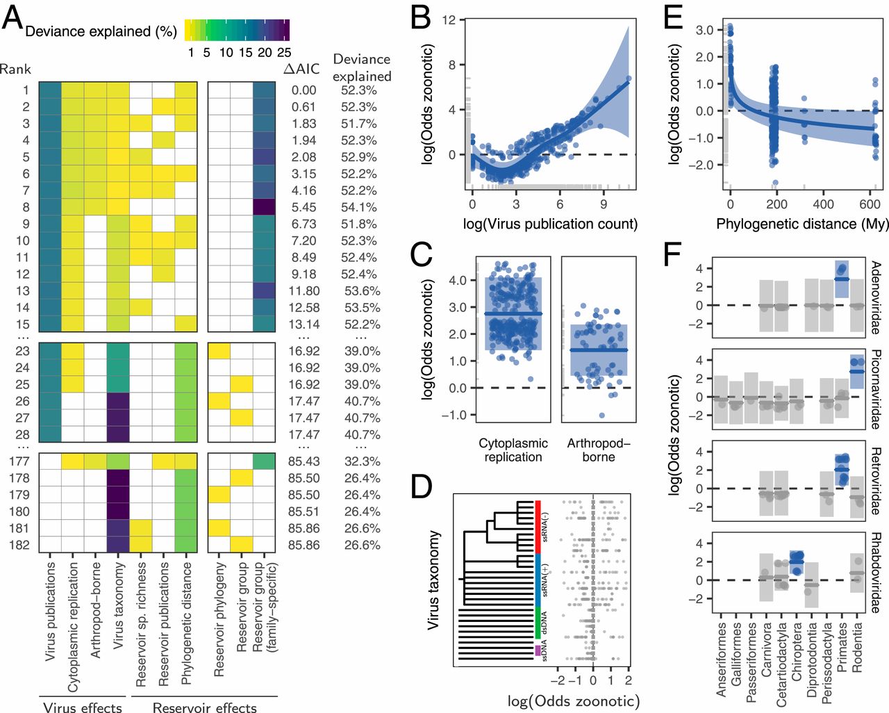
Among effects related to reservoir hosts, zoonotic risk declined with greater phylogenetic distance from reservoirs to primates, but this effect explained only 0.7% of the deviance in the top model and was absent from other competitive models (Fig. 2 A and E). Effects of reservoir order or phylogeny (either alone or crossed with an effect of virus family-level taxonomy) did not occur until the 23rd-ranked model, where they explained <0.01% of the deviance (ΔAIC from top model >16.9; Fig. 2A). The only strongly supported models containing an effect of reservoir host restrained the effect to certain viral families (i.e., a random effect of reservoir nested within virus family, here termed the “virus family-specific reservoir” effect). This effect explained 20.7% of the deviance in zoonotic status in the top model. In particular, primate adenoviruses and retroviruses, bat rhabdoviruses, and rodent picornaviruses were more likely to be zoonotic relative to viruses from the same families associated with other reservoirs (Fig. 2F). Critically however, none of these reservoir groups increased zoonotic propensity across additional viral families, suggesting the absence of generalizable effects of reservoir group on the likelihood of zoonotic transmission (Fig. 2A and SI Appendix, Fig. S2).
We further evaluated whether effects of reservoir hosts on zoonotic status might have been missed due to the inclusion of potentially collinear variables or insufficient statistical power to fit complex models. First, given that study effort understandably increased for zoonotic viruses, including this factor might have disguised true reservoir effects. However, models which did not adjust for virus-related publication counts failed to support reservoir effects; the best of such models achieved similar AIC values with and without reservoir effects (models 177 to 182, Fig. 2A). Second, including virus taxonomy might have reduced the power of our models. However, reservoir effects remained negligible or absent in models which excluded viral taxonomy (SI Appendix, Fig. S3). In contrast, viral family effects were robust to inclusion of general reservoir effects (i.e., those testing for consistent reservoir effects across viral families). When the family-specific reservoir effect was not present, viral rather than reservoir taxonomy was the stronger predictor of zoonotic status (models 23 to 28 in Fig. 2A and SI Appendix, Fig. S3). Thus, our results provide no support for the special reservoir hypothesis, which predicted that viruses associated with certain reservoir groups would be more likely to be zoonotic.
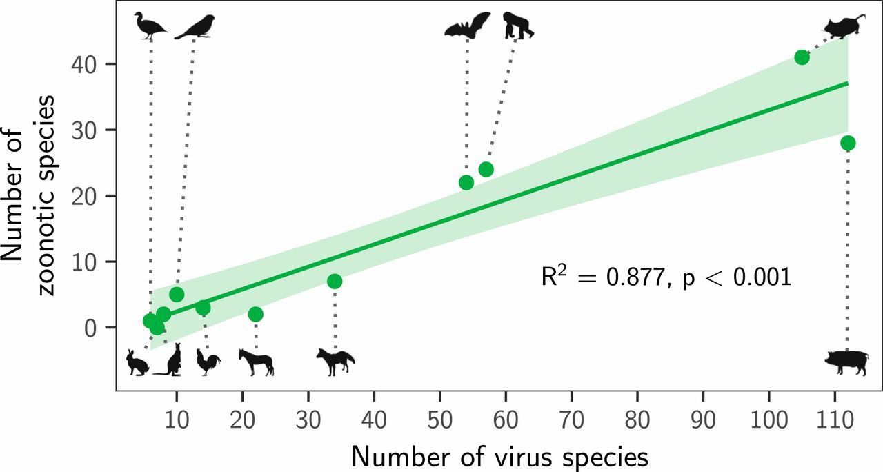
Instead, both the total number of viruses associated with each reservoir group (“viral richness”) and the number of zoonoses matched expectations under the reservoir richness hypothesis. The absence of reservoir effects meant that viral richness was strongly correlated with the number of zoonotic species (R2 = 0.88, P < 0.001; Fig. 3), consistent with a similar per-virus zoonotic risk across reservoirs. Notably, a similar correlation was previously observed when measuring viral richness at the host species level (9). A series of negative-binomial generalized linear models showed that—after controlling for greater viral richness in mammals compared to birds—reservoir species richness was correlated with both the number of zoonotic viruses (65.5% of deviance) and the viral richness (56.4% of deviance) of different reservoir orders (Fig. 4 and SI Appendix, Fig. S4). These models outcompeted those including previously identified effects of virus-related research effort and the phylogenetic distance of each reservoir group from primates (9), which when included explained a maximum of 3.1% additional deviance (ΔAIC ≥ 1.09; models 2 to 4 in Fig. 4A). Exchanging the reservoir class effect (i.e., birds versus mammals) for phylogenetic distance to primates performed considerably worse than the top model (ΔAIC = 4.72; model 5 in Fig. 4A). Similar results were observed for models predicting total viral richness (SI Appendix, Fig. S4).
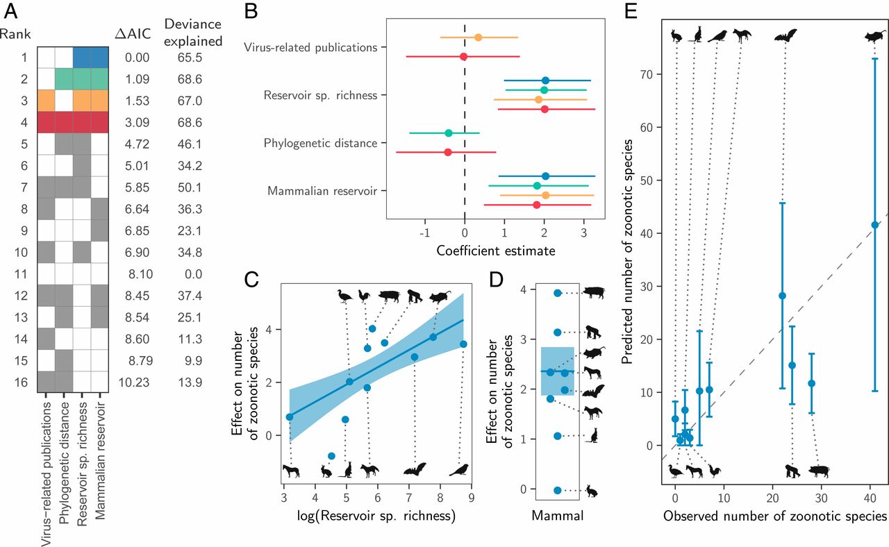
We next assessed how well the predictions of the reservoir richness model matched observations in individual reservoir groups. Highly recognized mammalian reservoir groups including bats and rodents hosted close to the number of zoonoses expected from their species richness (bats: 22 observed and 28 predicted [95% CI: 10.7 to 45.7]; rodents: 41 observed and 42 predicted [95% CI: 10.2 to 72.9]; Fig. 4E). In contrast, lagomorphs (rabbits, hares, and pikas) and diprotodonts (an order of marsupials) hosted both fewer viruses and fewer zoonoses than predicted; however, these were among the least-studied animal groups (Fig. 4E and SI Appendix, Fig. S4E). The only potentially “special” reservoirs identified by this analysis were the cetartiodactyls (even-toed ungulates and whales) and primates, which hosted more zoonoses than would be predicted for mammalian groups with their species richness (Fig. 4E). In the case of primates, this likely reflects the higher zoonotic propensity of primate-borne adeno- and retroviruses identified above (24 zoonotic viruses observed, of which 3 are adenoviruses and 7 are retroviruses; 15 predicted [95% CI: 7.8 to 22.4]). The mismatch was greater for cetartiodactyls (28 observed, 12 predicted [95% CI: 6.1 to 17.3]). However, perhaps due to their close association with humans as domestic livestock, this group also had considerably higher total viral richness than predicted (112 observed and 45 predicted [95% CI: 25.3 to 65.5]; SI Appendix, Fig. S4E). Consequently, the proportion of cetartiodactyl viruses which were zoonotic was unexceptional, inconsistent with the special reservoir hypothesis (Figs. 1 and 3). Strikingly, although mammals had elevated viral and zoonosis richness compared to birds, the proportion of zoonotic viruses was similar between classes, with 9/30 of avian and 115/387 of mammalian virus species being zoonotic (30% and 28.8%, respectively), emphasizing that—regardless of the cause—it is the underlying number of virus species which differs between mammals and birds, not the zoonotic component.
Animal groups have been proposed to differ in their propensity to transmit viruses to humans as a consequence of variation in their life history, physiology, or ecology. By combining a large dataset of reservoir host–virus associations with records describing their histories of human infection, our analysis found no evidence that the taxonomic identity of reservoirs affects the probability that the viruses they harbor are zoonotic. Instead, variation in the number of zoonoses maintained by each reservoir group was consistent with a largely host-neutral model, whereby more species-rich reservoir groups host more virus species and therefore a larger number of zoonotic species. These findings imply the need to reconsider some current approaches to virus discovery, surveillance, and research.
The absence of effects of reservoir host associations on the zoonotic status of viruses is at odds with the idea that conserved traits of certain host groups alter the zoonotic potential of their viruses. If host traits predictably altered zoonotic risk, these effects would be expected to act on multiple viral groups. Instead, we found that reservoir effects were isolated within individual virus families, such that no reservoir group altered the zoonotic risk of viruses across a broad range of viral families. For example, bats are widely considered special reservoirs due to their association with several high-profile zoonoses, including Severe acute respiratory syndrome-related coronavirus (Coronaviridae), Nipah henipavirus (Paramyxoviridae), and Ebola viruses (Filoviridae) (17, 19, 31). However, virus species within those families with a bat reservoir were no more likely to be zoonotic than those transmitted by other hosts, although we acknowledge that this effect would have been difficult to detect for filoviruses given the small number of virus species in this family. While our results do not dispute the existence of distinct features of bat immunity or life history which have been hypothesized to influence viral communities in bats (16, 18, 19, 32, 33), they provide no compelling evidence that these traits translate into an increased probability of bat-associated viruses infecting humans.
Idiosyncratic family-specific reservoir effects could reflect especially zoonotic clades within viral families that are strongly host-associated, sampling biases, or genuine interactions between the biological features of specific viral groups and specific reservoirs that increase zoonotic capability (e.g., differences in viral shedding or tissue tropism). Among rhabdoviruses, a significantly elevated proportion of bat-associated species were zoonotic, but this effect was driven by the rabies-causing genus Lyssavirus, which contained 73% of the 25 zoonotic rhabdovirus species and is strongly bat-associated (35). However, Rabies lyssavirus is also zoonotic when associated with carnivores and bat-associated Rhabdoviruses outside of the Lyssaviruses are not known to be zoonotic, suggesting that traits of this viral genus rather than its reservoirs are the underlying driver of the detected bat effect (SI Appendix, Fig. S3). The putatively heightened frequency of zoonotic rodent-associated picornaviruses might conceivably arise if transmission by rodents selects for viral traits that disproportionately promote zoonotic capability or if picornaviruses have similar zoonotic capability across reservoir groups but heightened ecological overlap favors zoonotic transmission from rodents. Yet, given that our dataset contained only three rodent-associated picornaviruses (all zoonotic) and primates and cetartiodactyls also hosted zoonotic picornaviruses (three out of four and one out of six zoonotic, respectively), a statistical artifact of low sample size cannot be ruled out. In contrast, effects of nonhuman primate reservoirs on the zoonotic potential of adenoviruses and retroviruses may have a biological basis. All primate-associated adenoviruses (three out of three) and 7 out of 12 primate-associated retroviruses were zoonotic, while none of the 15 adenoviruses and 16 retroviruses associated with other reservoirs have been reported to infect humans. This suggests that evolutionarily conserved similarities between nonhuman primates and humans may facilitate zoonotic transmission in these viral families. Yet, our results also highlight limitations of host phylogeny as a generalizable predictor of zoonotic capability since primate associations did not affect the zoonotic status in most of the viral families maintained by nonhuman primates. We hypothesize that the advantages conferred by phylogenetic relatedness are crucial for viral families that are inherently host-restricted (e.g., retroviruses, which must integrate into host genomes) but are less influential for viruses with less specialized infection and replication mechanisms. More generally, the relatively poor performance of phylogenetic distance in predicting either the proportion or the number of zoonoses from different reservoir groups (Figs. 2E and 4 A and B) suggests that the evolutionarily conserved factors that facilitate cross-species transmission within animal orders (25, 36, 37) are unlikely to extend over deeper evolutionary distances.
Our modeling framework allowed us to distinguish the relative contribution of host and virus features to zoonotic status. Consistent effects of viral taxonomy support the conclusion that zoonotic ability is a feature of viral clades rather than host associations (9, 28); however, the underlying viral trait determinants remain unresolved. As shown here (Fig. 2) and elsewhere, transmission by arthropod vectors and replication in the cytoplasm of host cells were linked to zoonotic transmission (9, 38). These findings are potentially explainable by selection for viral generalism arising from the need to replicate in both vertebrate and invertebrate hosts and the avoidance of the additional barriers associated with entering and replicating in the nucleus in novel host species, respectively. Yet, the site of viral replication within cells is invariant within viral families and arthropod-borne transmission is also strongly evolutionarily conserved (27, 39). Therefore, these traits have limited utility for explaining large variation in zoonotic risk within viral families. Identifying whether factors beyond ecological opportunity predict zoonotic status is vitally important to anticipate zoonotic transmission and to estimate the number of viruses which may emerge in the future.
The number of zoonoses scaled positively with the host species richness of animal orders (Fig. 4). Since reservoir species richness did not affect the probability that viruses were zoonotic (Fig. 2A), direct effects of species richness—which might occur if viral maintenance by more species rich host orders facilitates zoonotic transmission by selecting for broader host range—are unlikely (2). Instead, our results support a numerical, host species-neutral explanation: more species-rich animal orders maintain more viruses and hence more zoonoses. Indeed, after controlling for diminished viral richness in birds compared to mammals, key groups including bats and rodents harbored close to the number of zoonoses expected from their species richness (Fig. 4). These results mirror—and potentially explain—observations that the risk of zoonotic emergence is elevated in regions with high species richness (3, 40), since more virus species and hence zoonoses would be expected in species-rich habitats. More generally, our results suggest that the processes which shape viral richness (i.e., extinction, codivergence, and host shifts) do not vary enough among animal orders to create significant differences in the average number of viruses per species across reservoir orders. One implication of the relationship between reservoir species richness and the number of zoonoses is that surveillance efforts aimed at finding potential zoonoses should scale with the species richness of reservoir groups. This recommendation differs from the current practice which is based on the a priori expectation that certain groups (e.g., bats, rodents, and primates) are more likely to maintain zoonotic viruses (7). Analogously, our results underscore the challenges of identifying unknown reservoirs for viruses of importance to human health, since sampling effort would need to scale with local biodiversity, which itself may be uncertain.
Our dataset and analytical approach differed in several epidemiologically important ways from previous studies which suggested variation in viral zoonotic propensity across animal groups (9, 12). First, restricting virus–reservoir associations to the order level allowed us to test hypotheses on the role of broad animal groups on zoonotic origins in the face of widespread uncertainty in the identity of viral reservoirs at the host species level (22, 23, 27). This meant excluding human viruses that occasionally infect nonhumans and all associations between viruses and nonhuman hosts which are not currently considered to be important in natural transmission cycles. While it is conceivable that some excluded host–virus associations have an unrecognized role in transmission, adding 302 previously reported associations which did not meet our criteria did not qualitatively change our results (SI Appendix, Fig. S6). Second, we considered a single reservoir order for most viruses rather than modeling every association of each virus with each infected host species within that order independently. This was critical to avoid potentially spurious effects driven by surveillance effort and multiple observations of the same virus in closely related species. For example, Dengue virus (DENV) has been detected in at least 20 bat species, which comprise 76% of DENV–host associations (9, 12), but nonhuman primates, not bats, are the currently accepted reservoirs of zoonotic DENV outbreaks (41⇓–43). Our dataset therefore included a single primate reservoir since the majority of DENV–host associations would have obscured conclusions on zoonotic origins. Finally, unlike previous studies, we included avian viruses; however, restricting our analysis to mammalian viruses failed to recover reservoir host effects on zoonotic risk (SI Appendix, Fig. S7). One possible explanation for the apparent discrepancy between our findings and those based on shared virus detections could be that heightened surveillance for or susceptibility to zoonoses has led to elevated detections of spillover infections in previously identified “special hosts.” Our results highlight the importance of separating exposure and infection from transmission in future studies which involve reservoirs of infection.
A clear limitation of our approach was that we were unable to consider traits that vary within reservoir groups (e.g., reproductive rates, population size, and geographic range size) which may moderate the baseline zoonotic risk by altering viral richness, transmission dynamics, or ecological contacts of hosts with humans (44). In addition, even at the broad level of host taxonomy we used, knowledge of virus–reservoir relationships and zoonotic capability is incomplete and unevenly distributed among host groups (45). Analogous analyses of the capability of viruses to infect nonhuman hosts could also enable broader understanding of the determinants of cross-species transmission but will require constructing comprehensive datasets of infection histories in these alternative species. Finally, we evaluated only viral richness and whether viruses were reported to infect humans. Whether viruses from different reservoirs differ systematically in their pathogenicity, capacity to transmit among humans, or in their frequency of zoonotic transmission cannot be assessed from our data.
In summary, our analysis suggests that variation in the frequency of zoonoses among major bird and mammal reservoir groups is an emergent property of variation in host and virus species richness. We find no evidence that intrinsic or ecological differences among animal groups increases the number of viruses they maintain or the likelihood that any given virus is zoonotic. Basing public health surveillance and research strategies aiming to identify high-risk viruses on the assumption that some taxonomic orders of hosts are disproportionate sources of zoonoses risks missing important zoonotic viruses while simultaneously reenforcing patterns that may reflect detection biases rather than zoonotic risk.
We studied 35 virus families listed as infecting animals in either Fields Virology or the ViralZone web resource (Adenoviridae, Anelloviridae, Arenaviridae, Arteriviridae, Asfarviridae, Astroviridae, Birnaviridae, Bornaviridae, Caliciviridae, Circoviridae, Coronaviridae, Filoviridae, Flaviviridae, Genomoviridae, Hantaviridae, Hepadnaviridae, Hepeviridae, Herpesviridae, Nairoviridae, Orthomyxoviridae, Papillomaviridae, Paramyxoviridae, Parvoviridae, Peribunyaviridae, Phenuiviridae, Picobirnaviridae, Picornaviridae, Pneumoviridae, Polyomaviridae, Poxviridae, Reoviridae, Retroviridae, Rhabdoviridae, Tobaniviridae, and Togaviridae) (39, 46). For all data-collection steps below, virus names were matched to the latest accepted taxonomy according to version 2018b of the ICTV Master Species List by referring to historical taxonomic releases (47).
Following Babayan et al. (27), data on the reservoirs known to maintain the included virus species were obtained by searching common virology textbooks, such as Fields Virology (39), followed by in-depth literature searches until at least one reservoir or consistent evidence that the reservoir is currently considered as unknown was found. We summarized viral maintenance to the level of taxonomic orders of reservoirs because 1) the special reservoir hypothesis is generally articulated in terms of taxonomic order (e.g., applying to Chiroptera or Rodentia) and 2) order-level analyses constituted a reasonable compromise between taxonomic resolution and sample size, maximizing the number of virus species which could be included (27). We supplemented the single-stranded RNA virus dataset of Babayan et al. (27) with 344 additional records representing all recognized species in the families above for which reservoirs could be found. When there was evidence for independent maintenance by multiple reservoir orders, all known virus species–reservoir order combinations were recorded. For example, Rabies lyssavirus is maintained by both Carnivora and Chiroptera, but never as part of the same transmission cycle (48). Nineteen such virus species were detected, associated with 2.5 reservoir orders on average. Viruses known to be maintained by humans were retained if there was evidence for independent maintenance of a separate viral lineage in nonhuman hosts (n = 25) and otherwise discarded (n = 72). Yellow fever virus, for example, can be maintained by humans but also has an independent nonhuman primate reservoir community, and lineages from such maintenance cycles are zoonotic (39). Two virus species, Mammalian orthoreovirus and Usutu virus, had reservoir communities currently thought to span multiple taxonomic orders and were excluded from further analysis. Finally, the dataset was restricted to contain only viruses associated with mammalian and avian reservoir groups for which at least five viruses were known. The zoonotic status of individual virus species was obtained by combining records of detected human infections from Olival et al. (9) (n = 342 virus species) and Woolhouse and Brierley (29) (n = 70) with additional literature searching (n = 3). Only detections where the identity of the virus infecting humans was confirmed to species level by PCR or sequencing were considered.
We used logistic regression models to assess the association between reservoir group and zoonotic status (a binary response variable). Because at least one previously reported predictor of zoonotic status—publication count—was expected to be nonlinear (9), we used generalized additive mixed models (48). Three possible representations of a reservoir host effect were included (described below in more detail) and combined with all possible combinations of additional variables that were previously reported to predict zoonotic status. These included measures of research effort, the phylogenetic distance between each reservoir taxonomic order and primates, the species richness of each reservoir order, whether or not the virus was transmitted by arthropods or replicated exclusively in the cytoplasm, and a hierarchical representation of virus taxonomy. Previously reported variables relating to host species, such as geographic overlap with humans, could not be included when summarizing reservoirs at the level of taxonomic order.
The simplest reservoir effect was a random intercept for each reservoir taxonomic order, with all reservoir orders assumed independent. This represented the typically assumed special reservoir effect, in which some reservoirs are more prone to maintain zoonotic viruses than others. A second representation allowed clustering on the reservoir phylogeny to represent the hypothesis that related reservoir groups have correlated associations with zoonotic viruses. For example, mammalian reservoirs may be associated with a larger fraction of zoonoses than avian reservoirs, or related mammalian clades may share phylogenetically conserved features which shape their association with zoonoses. This phylogenetic random effect was implemented as a multivariate normal distribution, where phylogenetic relationships among taxa determined the amount of correlation between randomly sampled intercepts for different reservoir taxonomic orders (51). The variance–covariance matrix of this distribution took the form σ2Aσ2A, where σ2σ2 is a variance parameter to be estimated and A is a known variance–covariance matrix derived from a phylogeny (51, 52). We used a composite time-scaled reservoir phylogeny representing the mean divergence dates for all clades estimated across multiple studies, as contained in the TimeTree database (51). Following Longdon et al. (36), this phylogeny was converted to a variance–covariance matrix by assuming a Brownian motion model of trait evolution, using the vcv.phylo function in the APE package in R version 3.5.1 (52, 53). The third representation of a reservoir effect allowed independent random intercepts for each combination of reservoir order and virus family (i.e., the random effect of reservoir order was nested within virus family). This represented the hypothesis that specific reservoirs influence the propensity for viruses to be zoonotic for only some virus families, or that the identity of special reservoirs differs between families.
We used two measures to correct for the effects of variation in research effort on observations of zoonotic transmission: the numbers of results matching PubMed Central queries related to 1) virus species and 2) reservoir group (on 1 November 2019). For each virus species, this query was “<taxid>[Organism:noexp],” where “<taxid>” was replaced by the NCBI Taxonomy ID corresponding to each virus species. Viruses with no entry in the NCBI taxonomy database were removed from all analyses (this affected Cercopithecine gammaherpesvirus 14 and Hare fibroma virus, neither of which have publicly available sequence data in GenBank, meaning their reported reservoir associations are questionable). To capture a measure of the virus-related research effort in each reservoir group, a set of queries of the form “<taxid>[Organism:noexp] AND virus” were constructed, replacing “<taxid>” with the NCBI Taxonomy ID of each taxonomic order in turn. For nonhuman primates, this query was modified to “(txid9443[Organism:noexp] NOT txid9606[Organism:noexp]) AND virus,” where “txid9443” is the Taxonomy ID for the order Primates, and “txid9606” is the Taxonomy ID specific to Homo sapiens. Both variables were included as the natural logarithm of the respective publication counts, reflecting our prior belief that the effect of increasing numbers of publications would become saturated at high values. Nevertheless, we also allowed for additional nonlinear effects by fitting both variables as thin-plate smooths with 10 and 8 knots for virus and reservoir publication counts, respectively (54).
Phylogenetic distance was calculated as the cophenetic distance between each reservoir order and primates, using the same time-scaled reservoir phylogeny as above. Following Olival et al. (9), this effect was log(x + 1)-transformed in all models. The species richness represented by each reservoir order was derived from the Catalogue of Life using version 0.9.6 of the taxize library in R (55, 56). Both variables were fit as a thin-plate smooths with 6 and 10 knots, respectively.
The taxonomic random effect of virus family used the same specification as the reservoir phylogenetic effect above. Because the included virus families were too diverse to be represented in a single phylogeny (and might not share a common ancestor), a variance–covariance matrix representing currently accepted virus taxonomic relationships was generated. This matrix reflects the proportion of the taxonomy that is shared between virus families. Specifically, pairs of viruses were assigned a similarity score N/max(N), where N is the number of taxonomic levels they share, and max(N) the total number of taxonomic levels. This calculation was performed considering all taxonomic levels from realm to family. Thus, viruses in the same virus family received a similarity value of 1, while viruses from different families in the same order (e.g., Bunyavirales) received a value of 0.8. Virus species sharing no taxonomic levels were treated as independent (similarity = 0). Missing taxonomic assignments were interpolated to retain a comparable number of levels across all viruses, ensuring that all viruses remained independent at all levels not supported by the official ICTV taxonomy. For example, since many viruses are not classified using suborders a gap between order and family was bridged by creating a new level, assigning each family to its own unique suborder. This approach generated several free-floating branches, which are assumed independent in our models, but this is consistent with current virus taxonomy (ICTV Master Species List 2018b, https://ictv.global/files/master-species-lists/; SI Appendix, Fig. S3).
All models were fit using restricted maximum likelihood in the mgcv library in R and subsequently ranked by AIC (57, 58). The validity of models was checked using both standard methods implemented in the mgcv library and by inspecting simulated residuals generated using the DHARMa library in R (59). To calculate the proportion of the deviance explained by each term, each model was compared to submodels fit in the absence of individual terms, fixing the values for the smoothing parameters of the remaining terms to those estimated in the full model. The proportion of the deviance explained was calculated as (Di−DF)/D0(Di−DF)/D0, where DiDi is the deviance of model i, DFDF is the deviance of the full model, and D0D0 is the deviance of an intercept-only model.
To investigate potential explanations for differences in either the number of virus species or the number of zoonotic species associated with each reservoir order, two independent sets of negative binomial generalized linear models (GLMs) were fit to the counts of each virus type using the MASS library in R (60). Both sets of models contained all possible combinations of variables describing the log number virus-related publications associated with each reservoir, log phylogenetic distance to primates, log species richness in each reservoir, and a binary variable describing whether the reservoir is mammalian. We considered simpler Poisson GLMs instead of the negative binomial GLMs, but these showed strong overdispersion in simulated residuals using the DHARMa library.
The distribution of viral diversity among reservoir groups was quantified by calculating profiles of normalized alpha diversity using the rdiversity library in R (30). These calculations incorporated a species-level taxonomic similarity matrix, reflecting the proportion of the taxonomy that is shared between virus species and calculated as described for the family-level similarity matrix above.
Data and code used in this manuscript are available at https://doi.org/10.5281/zenodo.3516613.
We thank Dan Haydon and Roman Biek for helpful comments on earlier versions of this manuscript, as well as two anonymous reviewers, whose suggestions greatly improved the clarity of this publication. D.G.S. was supported by a Sir Henry Dale Fellowship, jointly funded by the Wellcome Trust and Royal Society (102507/Z/13/Z) and a Wellcome Senior Research Fellowship (217221/Z/19/Z). Additional funding was provided by the Medical Research Council through programme grants MC_UU_12014/8 and MC_UU_12014/12. Reservoir silhouettes were obtained from phylopic.org and were created by Matt Martyniuk (Anseriformes), Brian Gratwicke and T. Michael Keesey (Carnivora), and Sarah Werning (Lagomorpha), used with permission (https://creativecommons.org/licenses/by/3.0). Additional silhouettes by Andrew Butko (Passeriformes; https://creativecommons.org/licenses/by-sa/3.0) and Tamara L. Clark (Perissodactyla; https://creativecommons.org/licenses/by-nc-sa/3.0). All other silhouettes are in the public domain.
This open access article is distributed under Creative Commons Attribution License 4.0 (CC BY).
Viral zoonotic risk is homogenous among taxonomic orders of mammalian and avian reservoir hosts | PNAS
, Risk factors for human disease emergence. Philos. Trans. R. Soc. Lond. B Biol. Sci. 356, 983–989 (2001).OpenUrlCrossRefPubMedGoogle Scholar
, Host range and emerging and reemerging pathogens. Emerg. Infect. Dis. 11, 1842–1847 (2005).OpenUrlCrossRefPubMedGoogle Scholar
., Global trends in emerging infectious diseases. Nature 451, 990–993 (2008).OpenUrlCrossRefPubMedGoogle Scholar
, Infectious diseases: Preparing for the future. Science 313, 1392–1393 (2006).OpenUrlAbstract/FREE Full TextGoogle Scholar
., Prediction and prevention of the next pandemic zoonosis. Lancet 380, 1956–1965 (2012).OpenUrlCrossRefPubMedGoogle Scholar
., The global virome project. Science 359, 872–874 (2018).OpenUrlAbstract/FREE Full TextGoogle Scholar
., Building a global atlas of zoonotic viruses. Bull. World Health Organ. 96, 292–294 (2018).OpenUrlCrossRefGoogle Scholar
, Ecological origins of novel human pathogens. Crit. Rev. Microbiol. 33, 231–242 (2007).OpenUrlCrossRefPubMedGoogle Scholar
., Host and viral traits predict zoonotic spillover from mammals. Nature 546, 646–650 (2017).OpenUrlCrossRefPubMedGoogle Scholar
., Phylogenetic host specificity and understanding parasite sharing in primates. Ecol. Lett. 15, 1370–1377 (2012).OpenUrlCrossRefPubMedGoogle Scholar
, Diseases of humans and their domestic mammals: Pathogen characteristics, host range and the risk of emergence. Philos. Trans. R. Soc. Lond. B Biol. Sci. 356, 991–999 (2001).OpenUrlCrossRefPubMedGoogle Scholar
., A comparison of bats and rodents as reservoirs of zoonotic viruses: Are bats special? Proc. Biol. Sci. 280, 20122753 (2013).OpenUrlCrossRefPubMedGoogle Scholar
, Global patterns of zoonotic disease in mammals. Trends Parasitol. 32, 565–577 (2016).OpenUrlGoogle Scholar
, Origins of major human infectious diseases. Nature 447, 279–283 (2007).OpenUrlCrossRefPubMedGoogle Scholar
., Comparative analysis of bat genomes provides insight into the evolution of flight and immunity. Science 339, 456–460 (2013).OpenUrlAbstract/FREE Full TextGoogle Scholar
., Bat flight and zoonotic viruses. Emerg. Infect. Dis. 20, 741–745 (2014).OpenUrlCrossRefPubMedGoogle Scholar
, Bats as ‘special’ reservoirs for emerging zoonotic pathogens. Trends Microbiol. 23, 172–180 (2015).OpenUrlCrossRefPubMedGoogle Scholar
., Contraction of the type I IFN locus and unusual constitutive expression of IFN-α in bats. Proc. Natl. Acad. Sci. U.S.A. 113, 2696–2701 (2016).OpenUrlAbstract/FREE Full TextGoogle Scholar
., Dampened NLRP3-mediated inflammation in bats and implications for a special viral reservoir host. Nat. Microbiol. 4, 789–799 (2019).OpenUrlGoogle Scholar
., Accelerated viral dynamics in bat cell lines, with implications for zoonotic emergence. eLife 9, e48401 (2020).OpenUrlGoogle Scholar
, “Are bats exceptional viral reservoirs?” in New Directions in Conservation Medicine: Applied Cases of Ecological Health, A. A. Aguirre, R. Ostfeld, P. Daszak, Eds. (Oxford University Press, 2012), pp. 195–212.Google Scholar
, Identifying reservoirs of infection: A conceptual and practical challenge. Emerg. Infect. Dis. 8, 1468–1473 (2002).OpenUrlCrossRefPubMedGoogle Scholar
., Assembling evidence for identifying reservoirs of infection. Trends Ecol. Evol. 29, 270–279 (2014).OpenUrlCrossRefPubMedGoogle Scholar
., Consensus statement: Virus taxonomy in the age of metagenomics. Nat. Rev. Microbiol. 15, 161–168 (2017).OpenUrlCrossRefPubMedGoogle Scholar
., Host phylogeny constrains cross-species emergence and establishment of rabies virus in bats. Science 329, 676–679 (2010).OpenUrlAbstract/FREE Full TextGoogle Scholar
, Family level phylogenies reveal modes of macroevolution in RNA viruses. Proc. Natl. Acad. Sci. U.S.A. 108, 238–243 (2011).OpenUrlAbstract/FREE Full TextGoogle Scholar
, Predicting reservoir hosts and arthropod vectors from evolutionary signatures in RNA virus genomes. Science 362, 577–580 (2018).OpenUrlAbstract/FREE Full TextGoogle Scholar
., Taxonomic patterns in the zoonotic potential of mammalian viruses. PeerJ 6, e5979 (2018).OpenUrlCrossRefGoogle Scholar
, Epidemiological characteristics of human-infective RNA viruses. Sci. Data 5, 180017 (2018).OpenUrlGoogle Scholar
., How to partition diversity. arXiv:14046520v3 (8 December 2016).Google Scholar
, binom: Binomial confidence intervals for several parameterizations (R package, Version 1.1-1, 2014).Google Scholar
, Bats: Important reservoir hosts of emerging viruses. Clin. Microbiol. Rev. 19, 531–545 (2006).OpenUrlAbstract/FREE Full TextGoogle Scholar
., Network analysis of host-virus communities in bats and rodents reveals determinants of cross-species transmission. Ecol. Lett. 18, 1153–1162 (2015).OpenUrlCrossRefGoogle Scholar
., MHC class II proteins mediate cross-species entry of bat influenza viruses. Nature 567, 109–112 (2019).OpenUrlCrossRefGoogle Scholar
, Lyssaviruses and rabies: Current conundrums, concerns, contradictions and controversies. F1000 Res. 6, 184 (2017).OpenUrlGoogle Scholar
, Host phylogeny determines viral persistence and replication in novel hosts. PLoS Pathog. 7, e1002260 (2011).OpenUrlCrossRefPubMedGoogle Scholar
, Phylogenetically related and ecologically similar carnivores harbour similar parasite assemblages. J. Anim. Ecol. 83, 671–680 (2014).OpenUrlGoogle Scholar
, Ability to replicate in the cytoplasm predicts zoonotic transmission of livestock viruses. J. Infect. Dis. 199, 565–568 (2009).OpenUrlCrossRefPubMedGoogle Scholar
, Eds., Fields Virology (Lippincott Williams & Wilkins, ed. 6, 2013).Google Scholar
., Global hotspots and correlates of emerging zoonotic diseases. Nat. Commun. 8, 1124 (2017).OpenUrlCrossRefPubMedGoogle Scholar
., Experimental infection of Artibeus intermedius bats with serotype-2 dengue virus. Comp. Immunol. Microbiol. Infect. Dis. 36, 193–198 (2013).OpenUrlCrossRefPubMedGoogle Scholar
., Experimental inoculation of Artibeus jamaicensis bats with dengue virus serotypes 1 or 4 showed no evidence of sustained replication. Am. J. Trop. Med. Hyg. 91, 1227–1234 (2014).OpenUrlAbstract/FREE Full TextGoogle Scholar
., Neotropical bats that co-habit with humans function as dead-end hosts for dengue virus. PLoS Negl. Trop. Dis. 11, e0005537 (2017).OpenUrlGoogle Scholar
, Rodent reservoirs of future zoonotic diseases. Proc. Natl. Acad. Sci. U.S.A. 112, 7039–7044 (2015).OpenUrlAbstract/FREE Full TextGoogle Scholar
, Using metagenomics to characterize an expanding virosphere. Cell 172, 1168–1172 (2018).OpenUrlCrossRefGoogle Scholar
, ViralZone. https://viralzone.expasy.org/. Accessed 21 October 2019.Google Scholar
., Virus taxonomy: The database of the international committee on taxonomy of viruses (ICTV). Nucleic Acids Res. 46, D708–D717 (2018).OpenUrlCrossRefGoogle Scholar
, The role of viral evolution in rabies host shifts and emergence. Curr. Opin. Virol. 8, 68–72 (2014).OpenUrlCrossRefGoogle Scholar
, Viral zoonotic risk is homogenous among taxonomic orders of mammalian and avian reservoir hosts (Version 1.0.0). Zenodo. http://doi.org/10.5281/zenodo.3516613. Deposited 23 October 2019.Google Scholar
, Fast stable restricted maximum likelihood and marginal likelihood estimation of semiparametric generalized linear models. J. R. Stat. Soc. B 73, 3–36 (2011).OpenUrlCrossRefPubMedGoogle Scholar
, General quantitative genetic methods for comparative biology: Phylogenies, taxonomies and multi-trait models for continuous and categorical characters. J. Evol. Biol. 23, 494–508 (2010).OpenUrlCrossRefPubMedGoogle Scholar
, Fast stable direct fitting and smoothness selection for generalized additive models. J. R. Stat. Soc. Series B Stat. Methodol. 70, 495–518 (2008).OpenUrlGoogle Scholar
, TimeTree: A resource for timelines, timetrees, and divergence times. Mol. Biol. Evol. 34, 1812–1819 (2017).OpenUrlCrossRefGoogle Scholar
, APE: Analyses of phylogenetics and evolution in R language. Bioinformatics 20, 289–290 (2004).OpenUrlCrossRefPubMedGoogle Scholar
, R: A Language and Environment for Statistical Computing (R Foundation for Statistical Computing, 2018).Google Scholar
, Thin-plate regression splines. J. R. Stat. Soc. B 65, 95–114 (2003).OpenUrlCrossRefGoogle Scholar
, Taxize: Taxonomic search and retrieval in R. F1000 Res. 2, 191 (2013).OpenUrlGoogle Scholar
., Eds., Species 2000 & ITIS Catalogue of Life, 2015 Annual Checklist (Naturalis, 2015).Google Scholar
, A new look at the statistical model identification. IEEE Trans. Automat. Contr. 19, 716–723 (1974).OpenUrlCrossRefGoogle Scholar
, Smoothing parameter and model selection for general smooth models. J. Am. Stat. Assoc. 111, 1548–1563 (2016).OpenUrlCrossRefGoogle Scholar
, DHARMa: Residual diagnostics for hierarchical (multi-level / mixed) regression models (R package, Version 0.2.4, 2019).Google Scholar
, Modern Applied Statistics with S (Springer, ed. 4, 2002).Google Scholar
04/09/2020
Merlin D. Tuttle
I am writing in response to the unprecedented situation now presented by COVID-19 in North America. Early suggestions regarding potential transmission of SARS-CoV2 from humans to bats have raised serious concerns among research colleagues, rehabilitators, and cavers. In perusing available knowledge, I agree with veterinarian, Renee Schott. She has pointed out that we can’t control the public and their interactions with bats. Also, she notes that “COVID is everywhere in the U.S. right now. If North American bats can get it, they will get it.” She further warns that inappropriate restrictions could hasten the spread.
I sympathize with federal and state agencies who will be expected to take protective actions. Nevertheless, premature action could prove worse than none at all. SARS-CoV2 is already present worldwide, and bats appear highly resistant to coronaviruses. We seem to be considering rather serious actions to ward off threats not yet proven to exist and that may have serious unintended consequences.
Researchers unable to rapidly comply with new restrictions are already facing costly disruptions and termination of projects essential to conservation. Rehabilitators, forbidden to further assist bats, are reporting high-risk public actions. How would caving restrictions be enforced in state and federally managed tour caves, for example at Mammoth Cave or Carlsbad? And how could long-term restrictions on public access to other caves be enforced? What are the potential consequences of frightening the public with hypothesized disease transmission from mere breathing?
When faced with the choice of protecting bat benefits versus one’s family from potential death, the outcome is clear. Public overreaction to hypothetical threats of disease from bat droppings, or even bat breath, could prove disastrous, leading to intolerance and widespread killing of bats. Media speculation has already caused harm that may last for decades.
Regardless of presence in bats, new COVID strains in humans and other animals likely will appear. State and federal agencies, who have a responsibility to protect wildlife, cannot stop all interactions with wildlife, and bats are far from the only animals that can contract COVID. It’s already in cats and dogs.
As veterinarian, John Huckabee, has suggested for bat rehabilitators, the watchful eyes of researchers and cavers could prove invaluable, both in protecting bats from fearful humans and in providing early surveillance. We can only hope that government decision makers will consider the Hippocratic oath—first do no harm.
For further information on the COVID-19 pandemic and how it relates to bats, please read my editorial “A Viral Witch Hunt” published in the March 27 edition of Issues in Science and Technology.